PTE assessment
Three types of angiography for PTE assessment
There are three types of angiography for assessing whether a patient with a confirmed diagnosis of CTEPH is suitable for pulmonary thromboendarterectomy (PTE) surgery, also known as pulmonary endarterectomy (PEA).
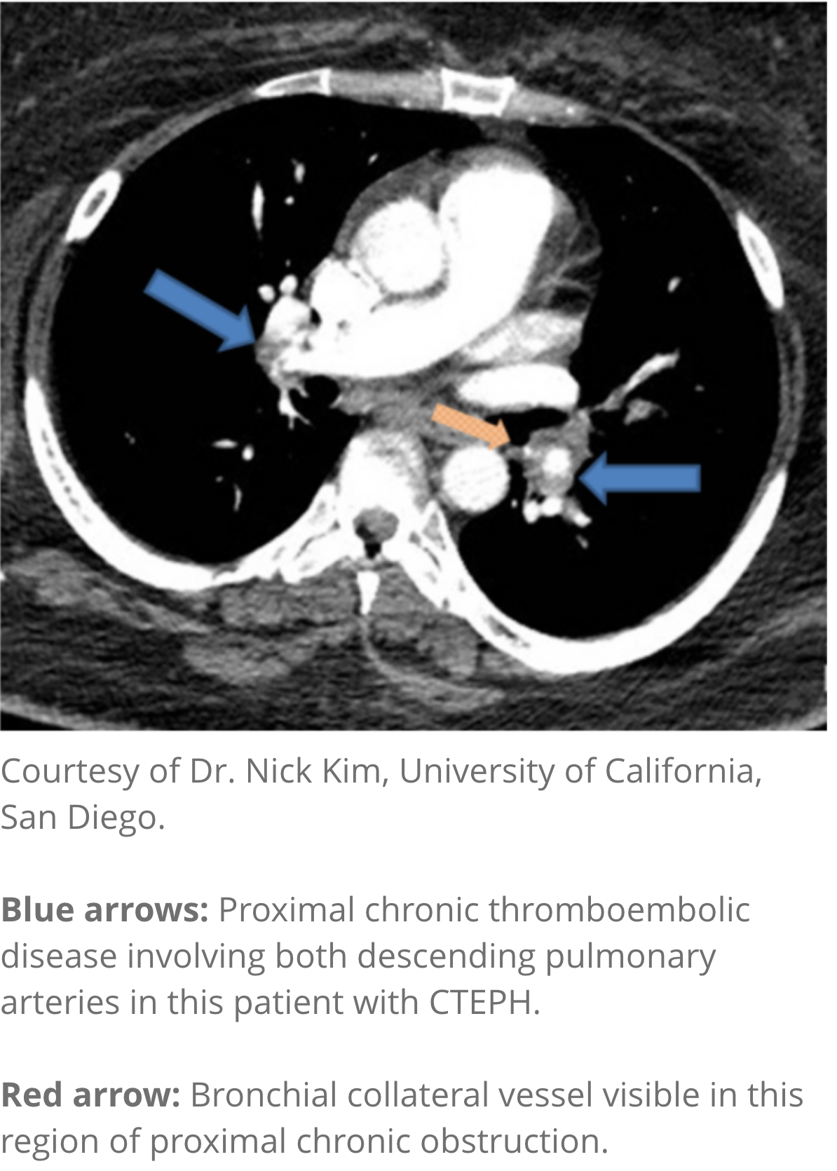
CT angiography
- Provides information regarding diagnosis and operability (eg, giving information on arterial walls)1,2
- Helpful in determining whether there is evidence of surgically accessible CTEPH1,2
- Note: Normal CT angiography does not exclude a diagnosis of CTEPH1,2

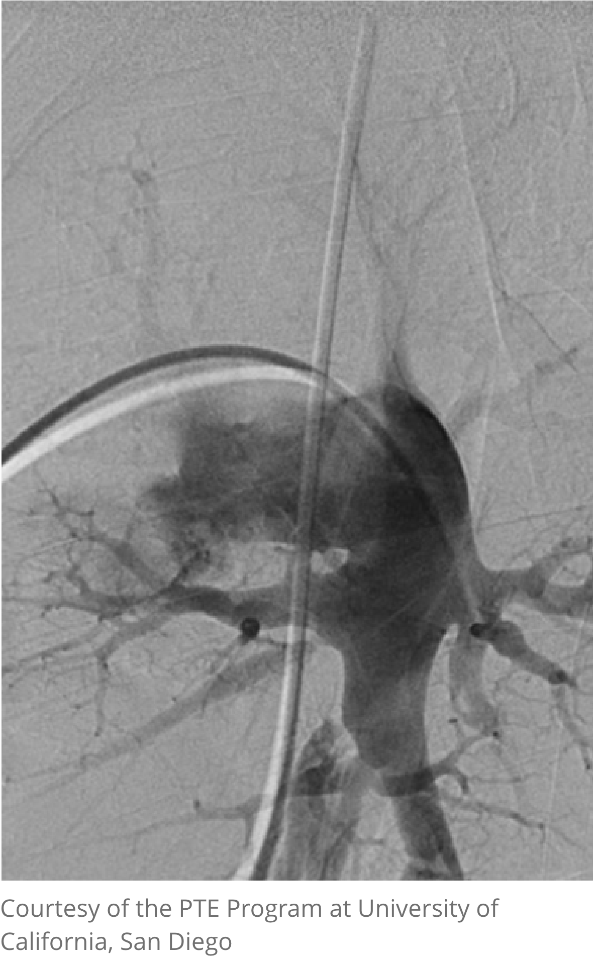
Pulmonary angiography
Pulmonary angiography (digital subtraction angiography) is used to assess treatment options when computed tomography pulmonary angiogram (CTPA) is inconclusive3
- Defines extent and distribution of disease and helps distinguish operable from inoperable disease4
- Combined with right heart catheterization (RHC), a correlation can be made between degree of disease and degree of hemodynamic impairment1,2
- The procedure should always be carried out by experienced staff at a unit with specialist pulmonary hypertension (PH) experience, preferably the unit at which PTE surgery would be performed1,2

MRI angiography
- Provides further information regarding diagnosis and operability, such as an evaluation of right-heart hemodynamics1,2
- Noninvasive technique does not involve exposure to radiation, so it is suitable for repeated studies2
- Note: Limited availability; may prove expensive and time consuming2
“A CTEPH team, consisting of an experienced PEA (PTE) surgeon and CTEPH physicians, should assess operability before alternative treatments are considered. Close working collaboration between community providers and CTEPH centers is required.”4
References:
1. Wilkens H, Lang I, Behr J, et al. Chronic thromboembolic pulmonary hypertension (CTEPH): updated recommendations of the Cologne Consensus Conference 2011. Int J Cardiol. 2011;154(suppl 1):s54-s60. 2. Jenkins D, Mayer E, Screaton N, Madani M. State-of-the-art chronic thromboembolic pulmonary hypertension diagnosis and management. Eur Respir Rev. 2012;21(123):32-39. 3. Humbert M, Kovacs G, Hoeper MM, et al. 2022 ESC/ERS Guidelines for the diagnosis and treatment of pulmonary hypertension. Eur Heart J. 2022;43(38):3618-3731. 4. Kim NH, D'Armini AM, Delcroix M, et al. Chronic thromboembolic pulmonary disease. J Am Coll Cardiol. 2013;62(suppl D):D92-D99.


